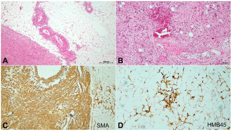
Figure 3. Photomicrographs of the kidney. A and B – Neoplastic tissue consisting of mature adipose tissue, with proliferation of smooth muscle tissue, newly formed vessels, and neoplastic cells with irregular nuclei increased of size (H&E original magnification 40x (A) and 100x (B)); C – Smooth muscle actin immunostaining confirmed the muscular component of the neoplasia (original magnification 40x); D – HMB45 immunohistochemistry staining was performed showing heterogeneous positivity in adipose cells, confirming the diagnosis of renal angiomyolipoma (original magnification 100x).