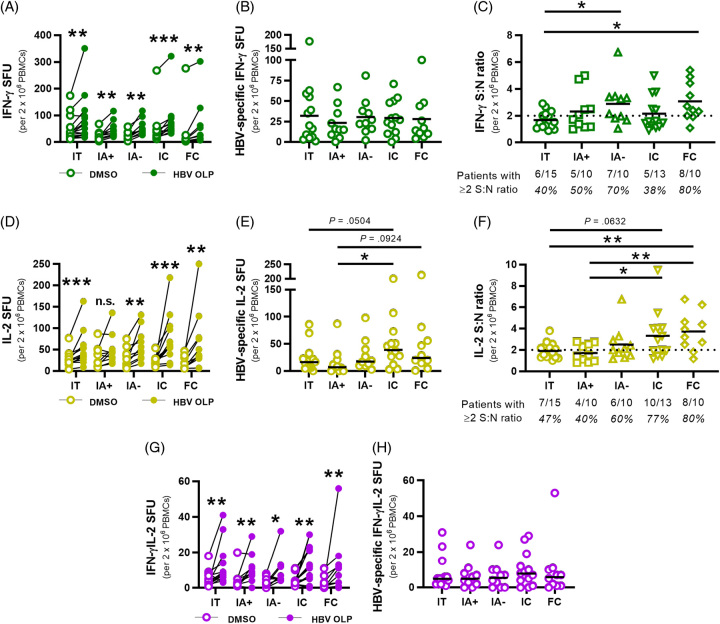
FIGURE 1.
Ex vivo detection of HBV-specific T cells in patient cohorts with chronic hepatitis B by means of multianalyte FluoroSpot assays. (A) IFN-γ+ SFU for HBV OLP (filled circles) and DMSO (empty circles) conditions were linked per patient in each cohort. (B) IFN-γ+ HBV-specific SFU for each patient were calculated by subtracting DMSO SFU from HBV OLP SFU. (C) IFN-γ+ signal:noise (S:N) ratios were calculated for each patient by dividing HBV OLP SFU by DMSO SFU. Ratios ≥2 were considered positive responses. Fractions beneath each cohort denote the number of patients with detectable responses. Black lines indicate sample means. (D) IL-2 + SFU for HBV OLP and DMSO conditions linked per patient in each cohort. (E) IL-2+ HBV-specific SFU for each patient from subtracting DMSO SFU from HBV OLP SFU. (F) IL-2+ S:N ratios for each patient by dividing HBV OLP SFU by DMSO SFU in each patient. (G) Multifunctional IFN-γ+IL-2+ SFU for HBV OLP and DMSO were linked per patient. (H) HBV-specific IFN-γ+IL-2+ SFU for each patient from subtracting DMSO SFU from HBV OLP SFU. Wilcoxon tests were conducted to compare SFU between HBV OLP and DMSO conditions (A, D, G). Mann-Whitney tests were used to compare HBV-specific SFU and S:N ratios between patient cohorts (B, C, E, F, H) (*p<0.05, **p<0.01, ***p<0.001). Abbreviations: FC, functionally cured; IA, immune-active; IC, inactive carrier; IFN-γ, interferon; IT, immunotolerant; OLP, overlapping peptide pool; PBMC, peripheral blood mononuclear cells; S:N ratio, signal-to-noise ratio; SFU, spot-forming unit.

