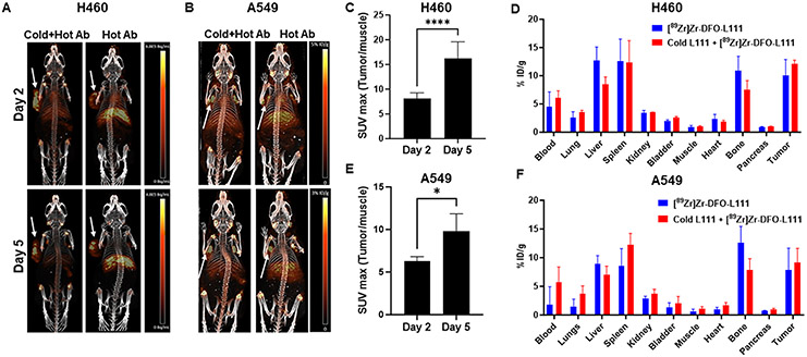
Figure 6. Small animal PET imaging and post-PET biodistribution.
A and B. PET imaging of the mice bearing subcutaneous H460 (A) and A549 (B) tumors (white arrows) injected with either [89Zr]Zr-DFO-L111 antibody or 4 mg/kg cold L111 prior to [89Zr]Zr-DFO-L111 antibody (n=4). Images at days 2 and 5 post-injection are shown. C. Bar graph showing the tumor-to-muscle SUVmax values at days 2 and 5 post-injection of the 4 mg/kg cold L111 prior to [89Zr]Zr-DFO-L111 antibody in H460 tumors. D. Post-PET biodistribution of the [89Zr]Zr-DFO-L111 antibody and the 4 mg/kg cold L111 injected prior to [89Zr]Zr-DFO-L111 antibody after five days post-injection in H460 tumor-bearing mice. E. Bar graph showing the tumor-to-muscle SUVmax values at days 2 and 5 post-injection of the 4 mg/kg cold L111 prior to [89Zr]Zr-DFO-L111 antibody in A549 tumors. D. Post-PET biodistribution of the [89Zr]Zr-DFO-L111 antibody and the 4 mg/kg cold L111 injected prior to [89Zr]Zr-DFO-L111 antibody after five days post-injection in A549 tumor-bearing mice.

