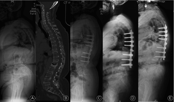
FIGURE 1.

A 65‐year‐old woman presented with severe back pain. (A). The standing long‐cassette lateral radiographs showed thoracolumbar hyper‐kyphosis arising from osteoporotic vertebral compression fracture (OVCF) and L5 spondylolisthesis, global kyphosis (GK) was 82.3°and thoracolumbar kyphosis (TLK) was 53.4°, sagittal stable vertebrae (SSV) located at L3. (B) Computed tomography revealed T9, T11, T12, and L1 fractures. (C) Postoperative lateral x‐ray showed T7‐L3 fixation and T11‐12 modified grade 4 osteotomy, GK decreased to 32.5°and TLK decreased to 6.2°, SSV moved cranially to L2. (D) Wedging in L34 disc was detected at 3‐years' follow‐up. (E) Degenerative changes progressed in L34 disc at 7‐years' follow‐up, GK and TLK was 55.1° and 10.6°, respectively.
