
FIGURE 3.
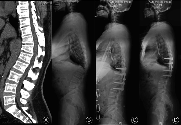
A 53‐year‐old man with degenerative thoracolumbar hyperkyphosis (DTH) arising from Scheuermann's kyphosis (SK). (A) Computed tomography showed wedging T11‐L2 vertebrae. (B) Lateral x‐ray showed global kyphosis (GK) and thoracolumbar kyphosis (TLK) were both 58.2°, sagittal stable vertebrae (SSV) located at L3. (C) Postoperative x‐ray showed fixation of two levels above and below the apex and modified grade 4 osteotomy, SSV moved cranially to L2, GK and TLK decreased to 21.9°. (D) Solid bony fusion could be seen without junctional complications at 2‐year follow‐up, GK and TLK maintained.