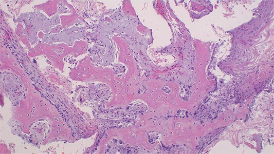
Figure 3.

Photomicroscopy of ossifying fibroma demonstrating a benign fibro-osseous nature of the lesion composed of diffuse hyperchromatic stromal fibroblastic cell proliferation, without atypia, or mitoses. The matrix is mineralized with woven and lamellar bone deposits or cementum-like calcifications distributed throughout the lesion, H & E staining at 200 × original magnification.
