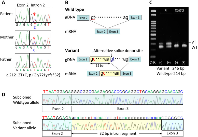
Fig. 2.
Summary of the molecular findings. A: Electrochromatograms showing a maternally derived splice donor site variant (c.212+2T>C) in intron 2 of GNAS-Gsα (marked with multiple asterisks) obtained using a forward primer hybridizing to exon 2 (5′-CTCTGCGTCGAAATGTCAAG-3′) and a reverse primer hybridizing to exon 3 (5′-TGGTTGCCTTCTCACCATC-3′). B: Schematic representation of the utilization of an alternative splice donor site and production of an aberrant mRNA predicted by SpliceAI. C: RT-PCR analysis of mRNA extracted from CHX-untreated and CHX-treated LCLs of the girl and a control subject. The primers utilized were: forward, 5′-GGGTGCTGGAGAATCTGGTA-3′; and reverse, 5′- TGGTTGCCTTCTCACCATC-3′. D: Electrochromatograms of subcloned wildtype and variant mRNA sequences obtained by RT-PCR for CHX-treated LCLs of the girl.

