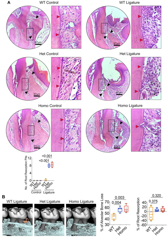
Figure 4.
IRF8 G388S promotes increased tooth root resorption and alveolar bone loss. (A) Histological analyses of maxillae (9-wk-old mice) by hematoxylin and eosin staining demonstrate increased tooth root resorption (red arrows) and alveolar bone loss (black arrows) in Irf8 KI Het and Homo mice compared to WT mice. Bar graph shows quantified root resorption pits. (B) Micro–computed tomography analyses of maxillae. Two-dimensional cut planes in sagittal orientation show alveolar bone loss around the ligated maxillary second molar. Scale bar: 0.5 mm. Bar graphs show quantified results for percentage of alveolar bone loss and tooth root resorption around the maxillary second molar in each group. Positive values indicate a loss in root structure/root resorption, while negative values will indicate minimal change or potentially an increase in root structure of ligated teeth. Panel A includes n = 4 mice per genotype (2 males and 2 females in each group; circle denotes male mice, and triangle denotes female mice). Panel B includes n = 5 to 6 mice per genotype (wild type [WT]: 3 males, 2 females; Het: 3 males, 3 females; Homo: 2 males, 3 females). The data are presented as the mean ± SD, and Het and Homo mice were compared against WT mice. One-way analysis of variance and post hoc Tukey’s test was used for comparisons among groups.

