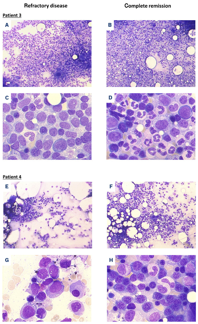
Figure 1.
Bone marrow cytomorpholog y. Bone marrow smears of patient 3 (A-D) and patient 4 (E-H) were analyzed by light microscopy after May-Grünwald and Giemsa staining at the time point of study enrollment (refractory disease after 1 cycle of cytarabine/daunorubicin therapy each) (A, C, E, G) and at the time point of complete response after the first cycle of study treatment (B, D, F, H); magnification 100x (A, B, E, F) and 630x oil immersion (C, D, G, H).

