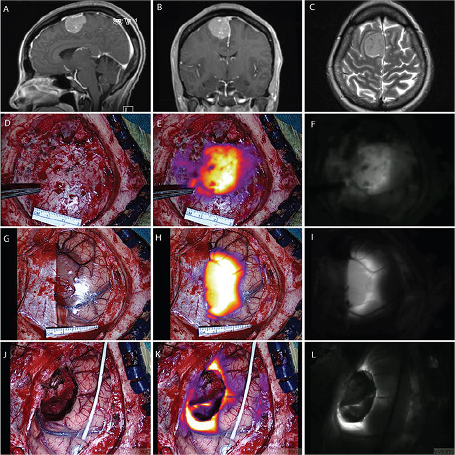
FIG. 1.

Case 1S. Convexity meningioma displaying NIR fluorescence. Sagittal (A) and coronal (B) Gd-enhanced T1-weighted MR images. Axial T2-weighted MR image (C) demonstrating minimal adjacent edema. Visible-light image (D), NIR fluorescence superimposed and color-mapped onto the visible-light view (E), and NIR image (F) showing closure of the dura, although some NIR signal can be seen through the dura and localized to tumor. Visible-light image (G), NIR fluorescence superimposed and color-mapped onto the visible-light view (H), and NIR image (I) showing dura reflected away and the tumor-brain interface. The SBR of NIR tumor signal is 6.05. Visible-light image (J), NIR fluorescence superimposed and color-mapped onto the visible-light view (K), and NIR image (L) showing residual fluorescence in anterior and posterior margins after resection of the main tumor. No fluorescence is seen in the midline falx.
