FIGURE 2.

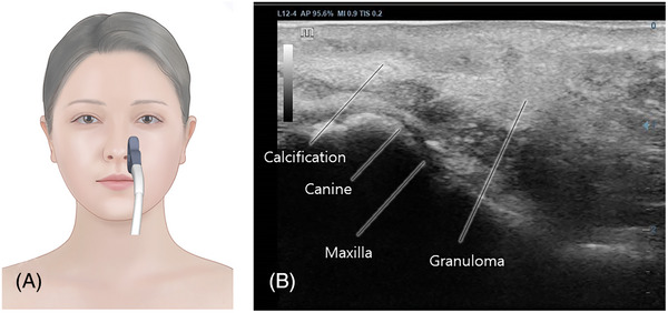
Ultrasonographic assessment of the left nasolabial fold in the longitudinal plane (A) depicted granulomas appearing as hyperechoic lesions beneath the dermal layer (B).

Ultrasonographic assessment of the left nasolabial fold in the longitudinal plane (A) depicted granulomas appearing as hyperechoic lesions beneath the dermal layer (B).