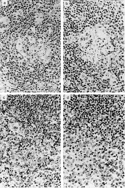
FIG. 4.
T-cell lymphoma in S. oedipus. Formalin-fixed tissue sections were stained with hematoxylin and eosin. (a and b) Germinal centers with follicular lysis. (c and d) Infiltration by a characteristic heterogeneous population of neoplastic lymphoid cells from T-cell areas with compression and loss of residual follicular B-cell areas. The photographs shown in panels a and c were obtained from C488 wild type-infected animals, and the photographs shown in panels b and d are from animals infected with ie14/vsag deletion mutant viruses. Original magnification, ×40.

