FIG. 4.
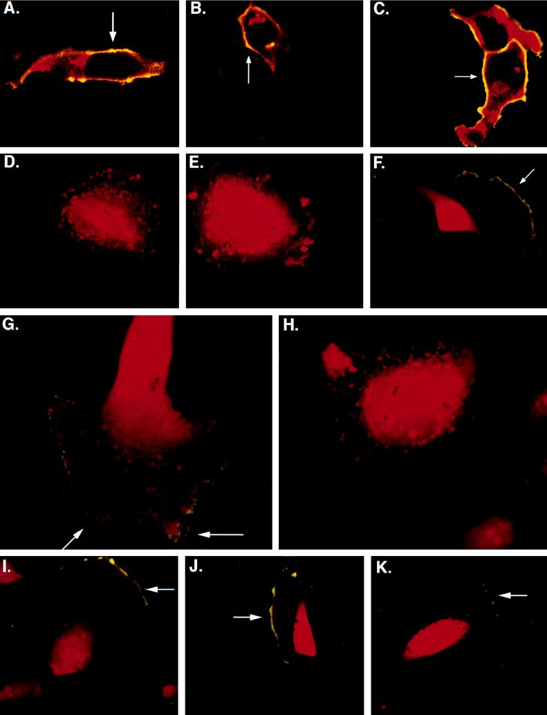
Subcellular localization of gB in HF and U373 cells. Confocal images of gB staining of cells infected with RVV gBwt, RVV gBAla, or RVV gBAsp were obtained. (A to C) Infected HF; (D to F) infected U373 cells. The cells were stained before permeabilization or at 4°C (representing surface gB) with mouse anti-gB (green) and after permeabilization (representing total gB) with mouse anti-gB (red). (A and D) RVV gBwt infections; (B and E) RVV gBAla mutant infections; (C and F) RVV gBAsp mutant infections. Surface gB staining is a combination of prepermeabilization at 4°C and postpermeabilization (yellow) (A, B, C, and F). (G and H) Confocal microscopy images demonstrating the presence of gB in VV-infected cells. U373 cells were infected with either RVV gBwt (G) or RVV gBAla (H) and subsequently treated with the phosphatase inhibitor tautomycin. The cells were stained with mouse antibody to gB prepermeabilization or at 4°C (green) (surface gB) and with mouse antibody to gB postpermeabilization (red) (total gB). The accumulation of gB trafficking to the surface was observed only in the RVV gBwt infection (yellow) (G). To demonstrate that transport of gB to the cell surface is not affected by the state of phosphorylation, we coinfected U373 cells with RVV gBwt (I), RVV gBAsp (J), or RVV gBAla (K) and RVV dynK44A. Magnifications, ×303 for panels A to F and I to K and ×473 for panels G and H.

