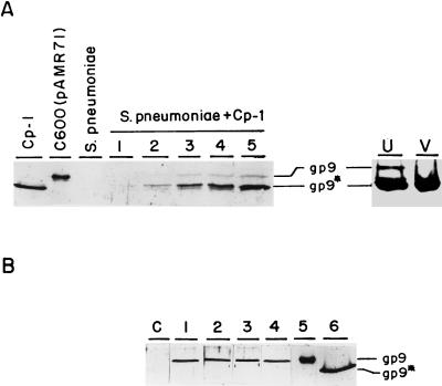
FIG. 2.
Western blot analysis of the major head protein of Cp-1 in different extracts. (A) Cp-1 corresponds to the Cp-1 virion; C600(pAMR71) extracts were prepared from E. coli; S. pneumoniae extracts were from uninfected cells; and lanes marked S. pneumoniae + Cp-1 contained extracts taken from Cp-1-infected cells at 40 (lane 1), 60 (lane 2), 80 (lane 3), 100 (lane 4), and 120 (lane 5) min after infection. On the right side are shown the results of analysis of the upper band (lane U) or the virion band (lane V) of a CsCl gradient of the Cp-1 lysate of S. pneumoniae (see text). (B) Extracts prepared from M31(pLSE1) (lane C), M31(pAMR71) (lane 1), UV-irradiated M31(pAMR71) (lane 2), mitomycin-treated M31(pAMR71) (lane 3), Dp-1-infected M31(pAMR71) (lane 4), C600(pAMR71) (lane 5), and the Cp-1 virion (lane 6). Samples were charged on an SDS–10% polyacrylamide gel and detected with a polyclonal anti-gp9* serum. The positions of gp9 and its processed form, gp9*, are indicated.

