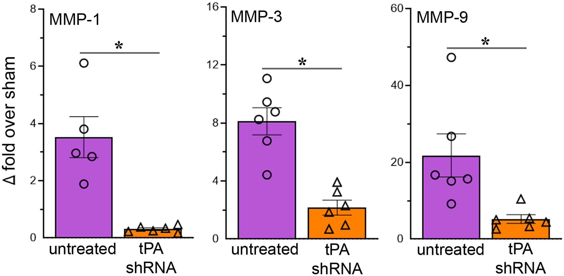
Fig. 3.

Reducing tPA after reperfusion decreases the expression of MMPs in the ischemic brain. The column scatter plots show the quantified mRNA expression (expressed as fold change over sham) of MMP-1, MMP-3, and MMP-9 in the ischemic brains of rats subjected to 2-h focal cerebral ischemia followed by reperfusion and euthanized on post-ischemic day 1. Columns indicate the mean and error bars indicate the SEM. *p < 0.05 (tPA shRNA vs untreated group).
