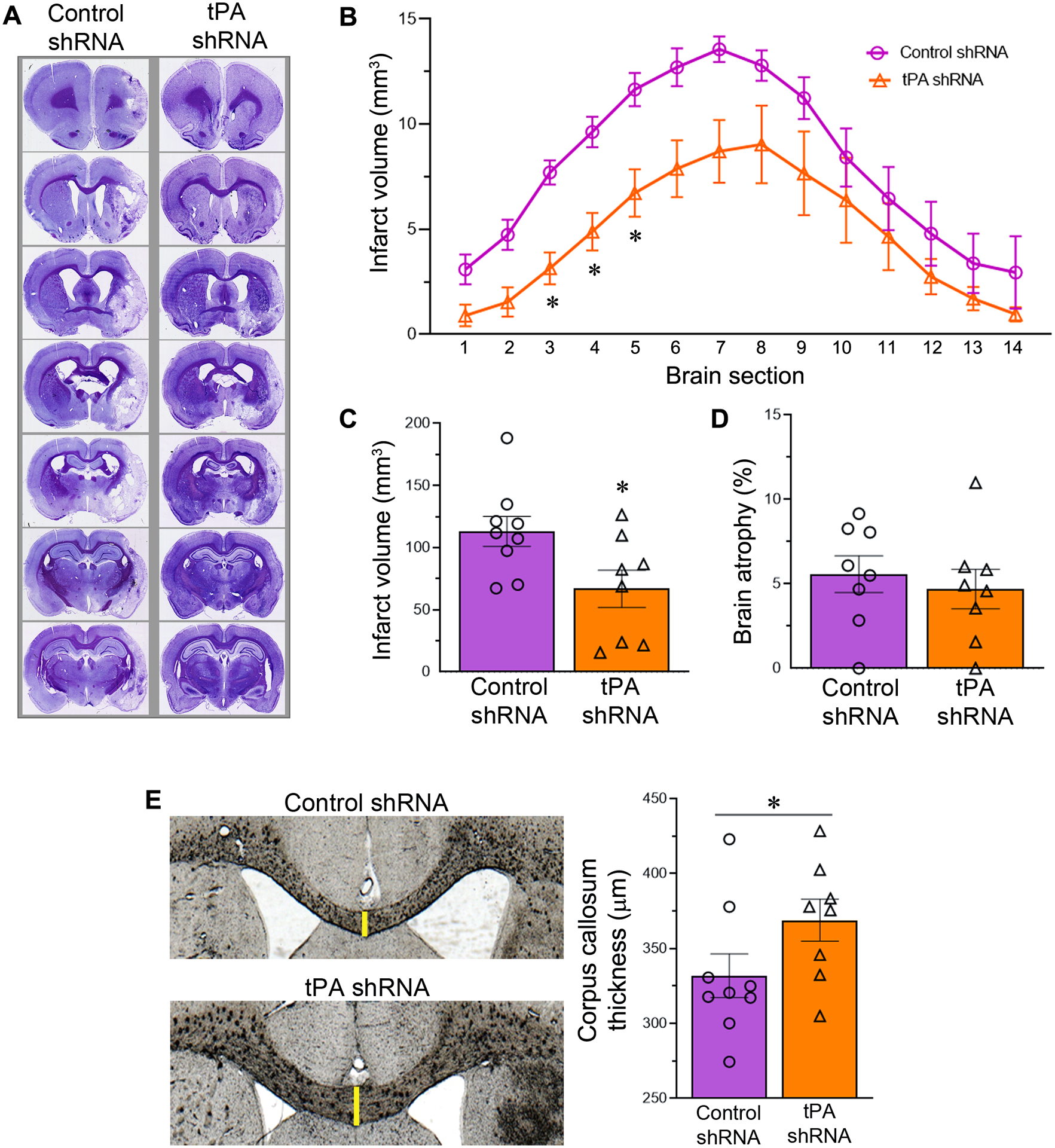
Fig. 5.

tPA shRNA treatment following transient focal cerebral ischemia and reperfusion attenuates infarct volume and white matter damage. (A) Representative cresyl violet stained coronal brain sections of control and tPA shRNA treated male rats subjected to 1.5-h focal cerebral ischemia followed by reperfusion for 14 days. The resulting stroke infarcts primarily affected the striatum and neocortex. (B) The line graph shows the quantified section by section infarct volume in both the experimental groups. n = 9 (control shRNA group) and 8 (tPA shRNA group). Error bars indicate the SEM. *p < 0.05 vs control shRNA group. (C) The column scatter plot shows the total infarct volume in both the experimental groups. Error bars indicate the SEM. *p < 0.05 vs control shRNA group. (D) The column scatter plot shows the brain atrophy in both the experimental groups. Error bars indicate the SEM. (E) Representative coronal brain sections depict the corpus callosum thickness in control and tPA shRNA treated male rats subjected to 1.5-h focal cerebral ischemia followed by reperfusion for 14 days. The column scatter plot shows the quantified corpus callosum thickness in both control shRNA and tPA shRNA treated rats. Columns indicate the mean and error bars indicate the SEM. *p < 0.05 (tPA shRNA group vs control shRNA group).
