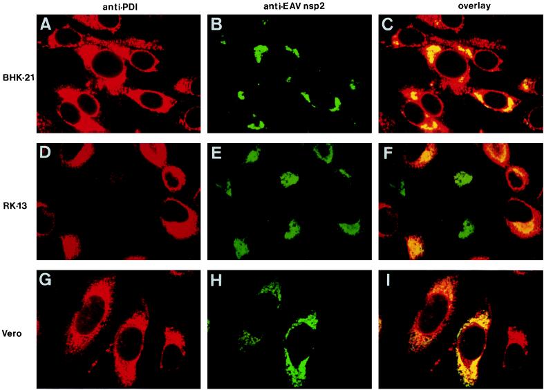
FIG. 3.
Colocalization of the EAV replicase and the cellular protein PDI, a marker for ER and IC. Three different cell lines were used: BHK-21 (A to C), RK-13 (D to F), and Vero (G to I). These were EAV infected, fixed at 8 h p.i., and processed for double-label immunofluorescence analysis with a mouse MAb directed against PDI (50), an anti-nsp2 rabbit antiserum (39), and appropriate secondary antibodies conjugated to fluorescent tags. PDI is shown in red (A, D, and G), and EAV nsp2 is shown in green (B, E, and H). For each sample, the differentially fluorescing images were recorded from the same optical section by using a confocal microscope. A computer-generated overlay of the PDI and EAV nsp2 images is shown (C, F, and I).

