Fig. 4:

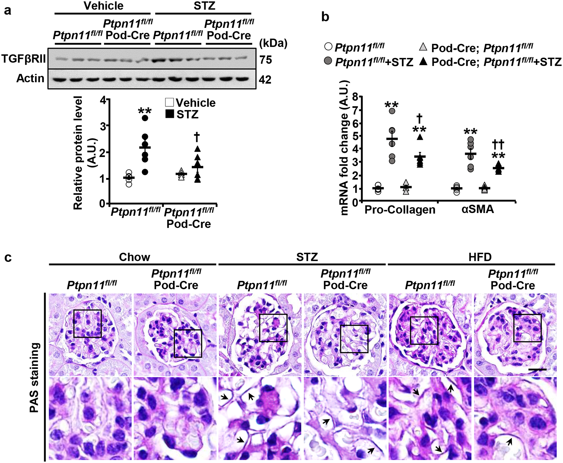
Podocyte Shp2 deficiency attenuates hyperglycemia-induced fibrosis in mice. a,b Immunoblots of TGFβRII and Actin (a), and mRNA expression of fibrosis markers pro-Collagen and αSMA (b) in kidneys from Ptpn11fl/fl and Ptpn11fl/fl; Pod-Cre mice treated without (vehicle) and with STZ (160 μg/g body weight, 20 weeks). Each lane in the immunoblot represents lysate from an individual animal. Actin served as a protein loading control for immunoblot (n=6 in each group), and mRNA expression was normalized to Tbp (n=6 in each group). *p ≤ 0.05, **p ≤ 0.01 vehicle versus STZ of mice with the same genotype. †p ≤ 0.05, ††p ≤ 0.01 Ptpn11fl/fl versus Ptpn11fl/fl; Pod-Cre under the same treatment. A.U.: arbitrary unit. c PAS staining of kidney sections from chow, STZ-treated (160 μg/g body weight, 20 weeks), and HFD-fed (24 weeks) Ptpn11fl/fl and Ptpn11fl/fl; Pod-Cre mice. Lower panel images are enlarged areas as highlighted by boxes. Arrows indicate glomerular basement membrane thickening. Scale bar: 20 μm.
