Fig. 5:

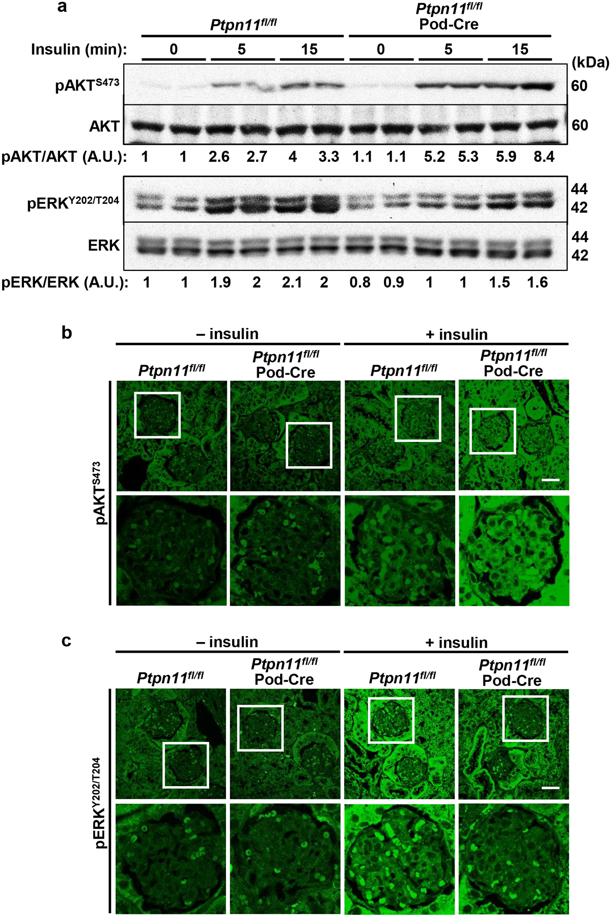
Podocyte Shp2 ablation enhances insulin-induced renal AKT signaling while attenuating ERK activation. a Immunoblots of pAKT S473, AKT, pERK Y202/T204, and ERK in kidney lysates from Ptpn11fl/fl and Ptpn11fl/fl; Pod-Cre mice without (0) and with insulin administration (10 U/kg body weight, for 5 and 15 minutes). Each lane represents lysate from an individual animal. Phosphorylation levels of AKT and ERK were normalized to the expression of the respective protein. A.U.: arbitrary unit. b, c Confocal images of kidney sections from Ptpn11fl/fl and Ptpn11fl/fl; Pod-Cre mice without (−) and with (+) insulin (15 minutes) immunostained for (b) pAKT S473 and (c) pERK Y202/T204. Boxed areas are enlarged in the lower panel. Scale bar: 25 μm.
