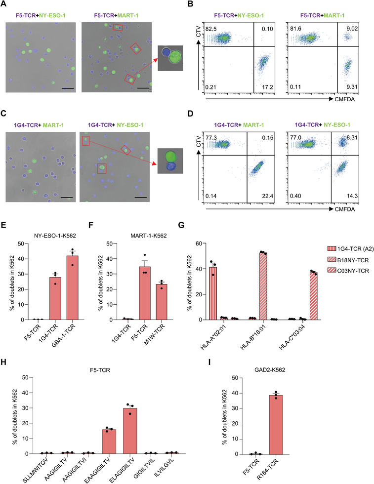
Figure 2.

DoubletSeeker serves as a powerful tool for the identification of interactions between the TCRs and pMHCs. A–D) Doublets formed by co‐incubation of CTV‐labeled F5‐TCR‐Jurkat (A,B) or 1G4‐TCR‐Jurkat (C,D) cells with CMFDA‐labeled NY‐ESO‐1‐K562 or MART‐1‐K562 cells (TCR‐expressing cells: SCT‐expressing cells = 5:1) were analyzed by confocal microscopy (A,C) and flow cytometry (B,D). F5‐TCR is paired with MART‐1‐SCT, and 1G4‐TCR is paired with NY‐ESO‐1‐SCT. Red rectangle boxes mark the presence of doublets. Scale bars, 50 µm. E) Percentage of doublets in NY‐ESO‐1‐K562 cells after co‐incubation with their cognate 1G4‐TCR‐Jurkat, GBA‐1‐TCR‐Jurkat or noncognate F5‐TCR‐Jurkat cells (TCR‐expressing cells: SCT‐expressing cells = 5:1) was analyzed by flow cytometry. F) Percentage of doublets in MART‐1‐K562 cells, following co‐incubation with Jurkat cells expressing the cognate F5‐TCR or M1W‐TCR (TCR‐expressing cells: SCT‐expressing cells = 5:1), was analyzed by flow cytometry. G) Antigen‐specific doublet formation between K562 expressing HLA‐A*02:01, HLA‐B*18:01 or HLA‐C*03:04 restricted NY‐ESO‐1 epitopes and Jurkat cells expressing their cognate TCRs (TCR‐expressing cells: SCT‐expressing cells = 5:1). H) Comparison of doublets formation in K562 cells expressing diverse HLA‐A*02:01‐restricted MART‐1 peptide variants after co‐incubation with F5‐TCR‐Jurkat cells. I) Percentage of doublets in K562 cells expressing MHC‐II restricted GAD2 antigen after co‐incubation with cognate R164‐TCR‐Jurkat or noncognate F5‐TCR‐Jurkat cells (TCR‐expressing cells: SCT‐expressing cells = 5:1) was analyzed by flow cytometry. CTV: CellTrace Violet, CMFDA: CellTrace CMFDA. Data are represented as the means ± SEM. N = 3. Data are representative of three independent experiments (A–I).
