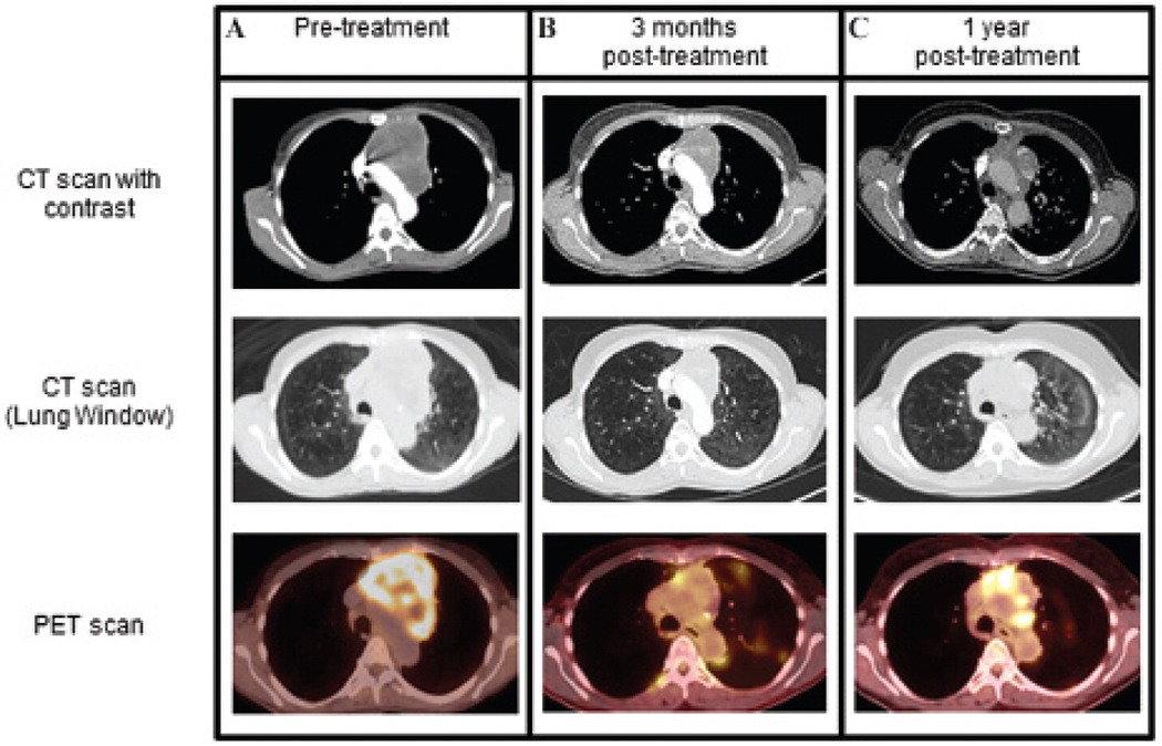
Figure 1.

Imaging presentation of an anterior mediastinal mass in patient D.H. A, On initial imaging, the patient presented with an anterior mediastinal mass with heterogeneous enhancement that measured 8.6 × 7.8 cm in size (upper CT image) (maximum SUV 10.7, lower [18F]FDG PET image). The great vessels were displaced medially from the left side, and the upper lobe branch of the left pulmonary artery was displaced inferolaterally. B, After chemoradiation, surveillance imaging demonstrated a reduction in the mass to 5.6 × 3.1 cm (upper CT image), with only a small rim of increased tracer uptake along the right medial and inferior margins and a maximal SUV of 6.1 (lower fusion [18F]FDG PET/CT image). Given the favorable response, further therapy was deferred. C, One year later, there was an increase in the [18F]FDG uptake in the medial and inferior aspects of the anterior mediastinal mass (SUV maximum 8.7, lower fusion [18F]FDG PET/CT image), suspicious for tumor recurrence.
