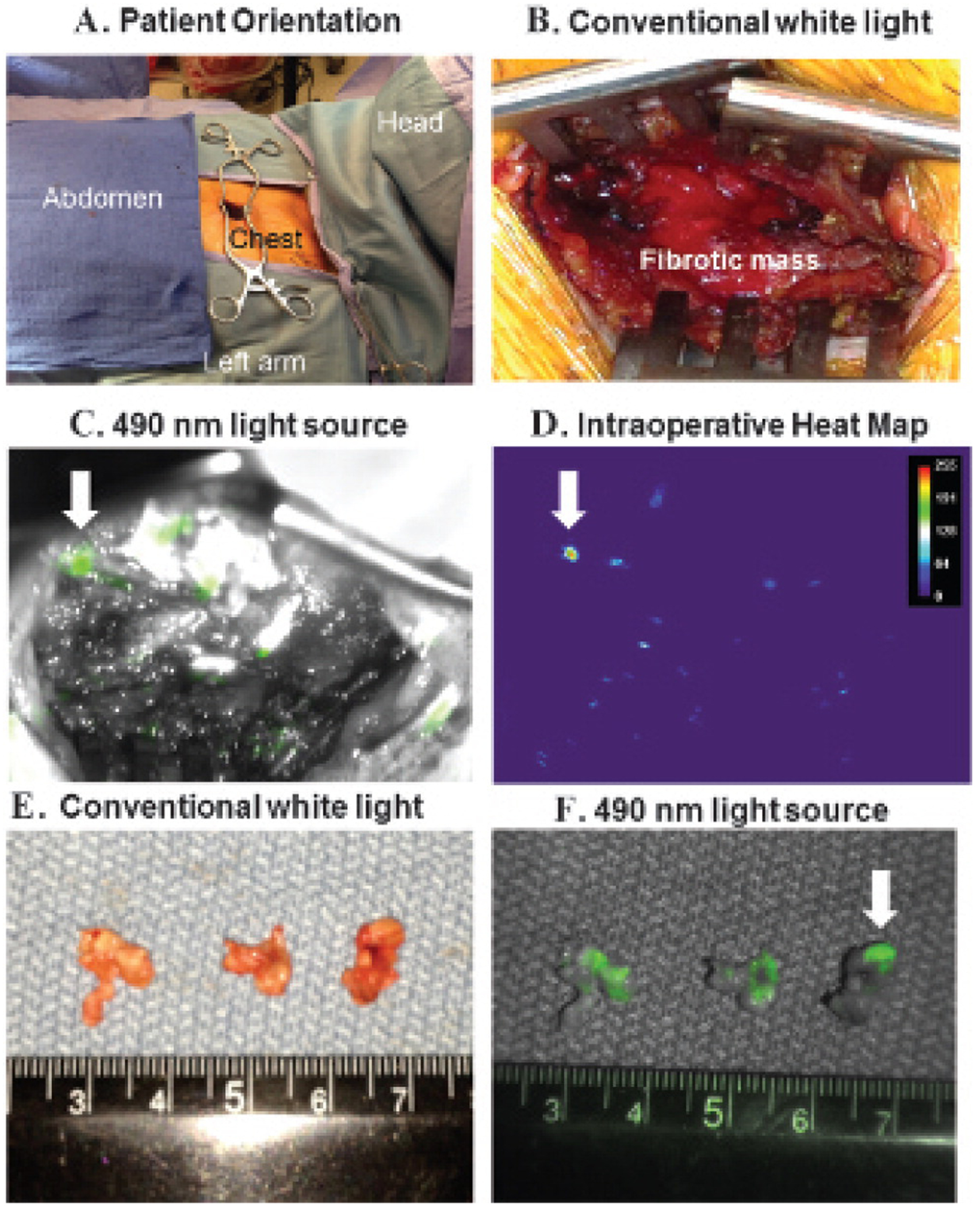
Figure 3.

Intraoperative images from the surgical biopsy of the anterior mediastinal mass. A, The patient was oriented with the head to the right and a 2 cm incision over the left chest just lateral to the left border of the sternum in the second intercostal space. B, After dissecting the pectoralis and intercostal muscles, the surgeon encountered a dense fibrotic mass that required a scalpel to penetrate. Multiple surgical biopsies did not reveal malignant cells. C, The incision was illuminated with a 490 nm light source and revealed several small foci of fluorescent deposits in the wound. The largest suspicious tumor deposit is marked with a white arrow. D, Heat map analysis confirmed several areas of the mediastinal mass that fluoresced from the targeted FOLR1 contrast agent. E, Three-subcentimeter tissue specimens were taken with a biopsy forceps. On gross inspection, the three specimens appeared homogeneous and not different from the previous biopsies. F, However, the specimens were reimaged ex vivo and fluoresced; thus, this gave the surgical team a high degree of confidence that it had located tumor cells in the chest.
