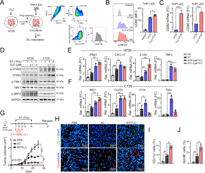
Fig. 3.
TLC388 synergistically enhanced RT-induced cancer immunogenicity. (A) The schematic diagram of immature DC cocultured with TLC388-treatment HT29 cell and analyzed DC maturation marker by flow cytometry. (B) The TLC388 (0.5 µM)-treated HT29 cells were cocultured with THP1-iDC cells, which were differentiated into immature DC (iDC) by IL-4 (1500 IU/ml) and GM-CSF (1500 IU/ml) for 7 days, for 24 h. The DC marker (CD86) was evaluated by flow cytometry. **p < 0.01. One-way ANOVA t-test. (C) HT29 cells were treated with TLC388 (0.5 µM) for 24 h, and then washout the drugs to coculture with THP1-iDC for 24 h. The mRNA level of DC markers (CD86 and CD80) was determined by qRT-PCR(n = 3). *p < 0.05 and **p < 0.01. One-Way ANOVA t-test. (D) HT29 and CT26 cells were irradiated for 5 Gy and treated with TLC388 (0.5 and 1 µM) for 24 h. The cell lysate was harvested for western blot analysis. (E) HT29 cells were irradiated with 5 Gy and treated with TLC388 (0.5 and 1 µM) for 24 h. The level of IFNβ1, CXCL10, IL12a, and TNFα mRNA was analyzed by qRT-PCR (n = 3). *p < 0.05 and **p < 0.01. One-way ANOVA t-test. (F) CT26 cells were irradiated with 5 Gy and treated with TLC388 (0.5 and 1 µM) for 24 h. The level of IFNβ1, CXCL10, IL12a, and TNFα mRNA was analyzed by qRT-PCR (n = 3). *p < 0.05 and **p < 0.01. One-way ANOVA t-test. (G) Tumor growth of CT26-driven colon carcinoma established in BALC/c mice (n = 6 per group) that were treated with RT (5 Gy for 2 fractions) and RT/TLC (TLC:2.5 mg/kg). Tumor growth is reported as the mean tumor volume ± SD. *p < 0.05 and **p < 0.01. CR: complete response. Two-way ANOVA t-test. (H) The tumor-infiltrating CD11c+ DC and GzmB+ immune cells within resected tumors were analyzed by immunofluorescent staining (n = 3). (I) The quantification of tumor-infiltrating CD11c+ dendritic cells within resected tumors (n = 3). *p < 0.05 and **p < 0.01. One-way ANOVA t-test. (J) The quantification of tumor-infiltrating GzmB+ T cells within resected tumors (n = 3). *p < 0.05 and **p < 0.01. One-way ANOVA t-test

