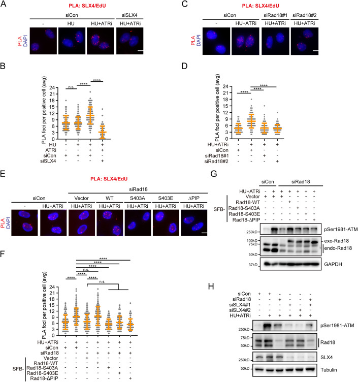
Figure 4. ATR restrains excessive SLX4 accumulation at stalled replication forks.
(A, B) HeLa cells were pulse-labeled with 10 μM EdU for 15 min, mock-treated, treated with 2 mM HU, or a combination of 2 mM HU and 2 μM VE-821 for 1 h, and then subjected to PLA with anti-SLX4 and anti-biotin antibodies. Representative images of PLA foci (red) (A). DNA was stained with DAPI. Scale bar, 10 μm. Quantification of PLA foci number per focus-positive cell (B). Data represent means ± SD of three independent experiments. More than 100 cells were counted for each sample. ****P < 0.0001, n.s., not significant, one-way ANOVA test. (C, D) Impaired SLX4 recruitment to stalled forks upon Rad18 depletion. HeLa cells were transfected with the indicated siRNAs, pulse-labeled with 10 μM EdU for 15 min, mock-treated or treated with a combination of 2 mM HU and 2 μM VE-821 for 1 h, and then subjected to PLA with anti-SLX4 and anti-biotin antibodies. Representative images of PLA foci (red) (C). DNA was stained with DAPI. Scale bar, 10 μm. Quantification of PLA foci number per focus-positive cell (D). Data represent means ± SD of three independent experiments. More than 100 cells were counted for each sample. ****P < 0.0001, n.s., not significant, one-way ANOVA test. (E, F) HeLa cells were transfected with the indicated siRNAs/plasmids, pulse-labeled with 10 μM EdU for 15 min, mock-treated or treated with a combination of 2 mM HU and 2 μM VE-821 for 1 h, and then subjected to PLA with anti-SLX4 and anti-biotin antibodies. Representative images of PLA foci (red) (E). DNA was stained with DAPI. Scale bar, 10 μm. Quantification of PLA foci number per focus-positive cell (F). Data represent means ± SD of three independent experiments. More than 100 cells were counted for each sample. ****P < 0.0001, n.s., not significant, one-way ANOVA test. (G) HeLa cells transfected with the indicated siRNAs/plasmids were mock-treated or treated with the combination of 2 mM HU and 2 μM VE-821 for 3 h. Immunoblotting was performed using antibodies as indicated. (H) HeLa cells were transfected with siRNAs as indicated. 48 h after transfection, cells were mock-treated or treated with a combination of 2 mM HU and 2 μM VE-821 for 3 h. Immunoblotting was performed using antibodies as indicated. Source data are available online for this figure.

