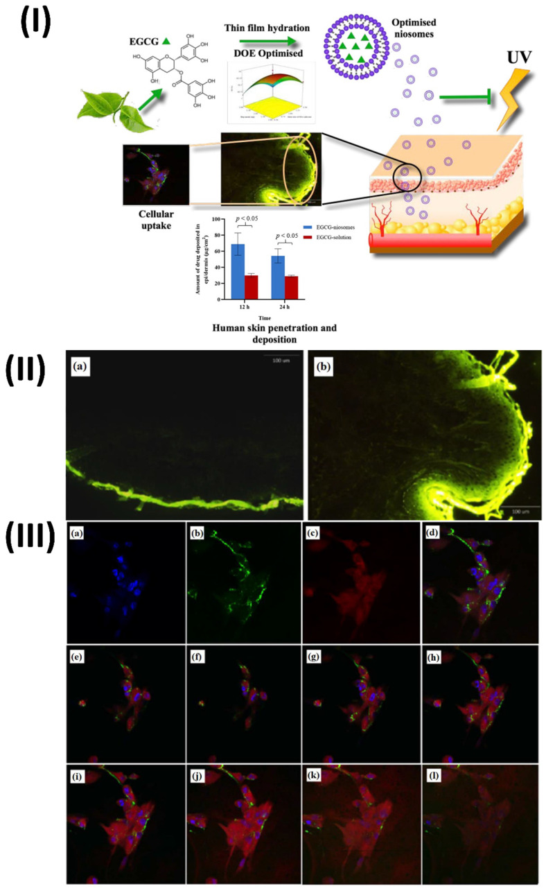
Figure 5.
(I) Technique of niosomes preparation and cellular mechanism (II) Sections of human skin treated with a FITC solution show the whole thickness of the skin. (III) Perinuclear particle accumulation is shown in confocal laser scanning microscopy pictures of Fbs following 2 hours of incubation with FITC-labelled nanosomes at 37 °C. Reproduced with the permission from ref. 80 Graphical abstract, Fig. 6, and Fig. 9 (MDPI).

