
FIG. 2.
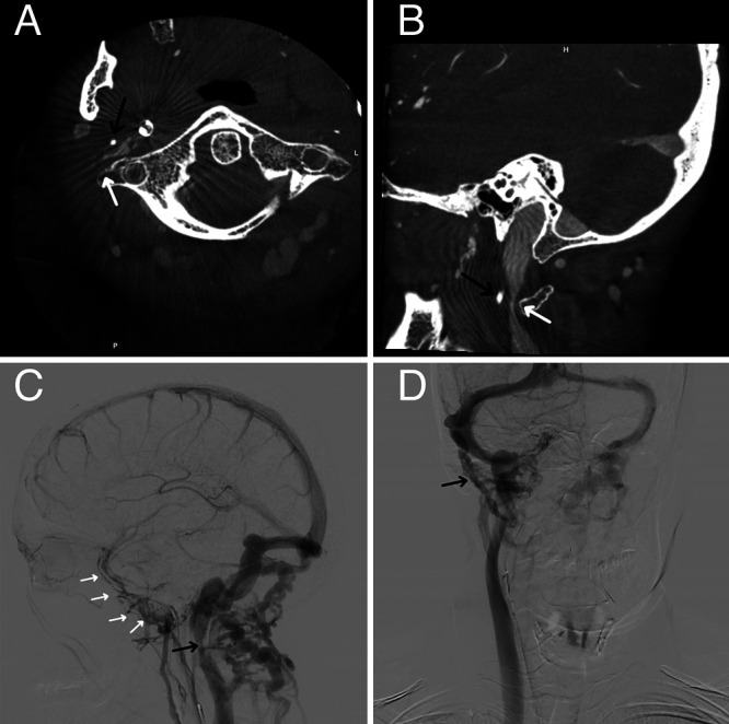
Axial (A) and sagittal (B) CT angiography showing impingement of the internal jugular vein between the styloid process (black arrows) and the arch of C1 (white arrows). Catheter angiography shows narrowing of the right internal jugular vein (black arrow, C) and reconstitution of flow to the right internal jugular vein immediately downstream to the presumed impingement point (white arrows, C) and dilated right-sided mastoid emissary vein (black arrow, D) and absence of blood flow on the left side.