. 2024 Feb 9;6(2):e230080. doi: 10.1148/rycan.230080

Figure 1:

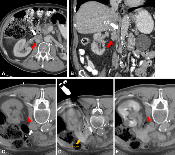
CT scans in a 67-year-old male patient with metastatic pheochromocytoma to the retroperitoneum. (A, B) Preprocedural CT scans demonstrate metastatic lymph node (red arrows, 2.4 × 2.0 × 3.7 cm) immediately posterior to the inferior vena cava (IVC; white arrows). (C) Patient in the prone treatment position demonstrating nodal target (red arrow). (D) One of two microwave ablation antennas in place after hydrodissection fluid was placed in the retroperitoneum (Hydro; yellow arrow). Treatment was performed for 5 minutes at 65 W. (E) Immediate postprocedural scan with intravenous contrast material. Note shrinkage of node (red arrow) after microwave ablation due to tissue dehydration.

CT scans in a 67-year-old male patient with metastatic pheochromocytoma to the retroperitoneum. (A, B) Preprocedural CT scans demonstrate metastatic lymph node (red arrows, 2.4 × 2.0 × 3.7 cm) immediately posterior to the inferior vena cava (IVC; white arrows). (C) Patient in the prone treatment position demonstrating nodal target (red arrow). (D) One of two microwave ablation antennas in place after hydrodissection fluid was placed in the retroperitoneum (Hydro; yellow arrow). Treatment was performed for 5 minutes at 65 W. (E) Immediate postprocedural scan with intravenous contrast material. Note shrinkage of node (red arrow) after microwave ablation due to tissue dehydration.