. 2024 Feb 9;6(2):e230080. doi: 10.1148/rycan.230080

Figure 3:

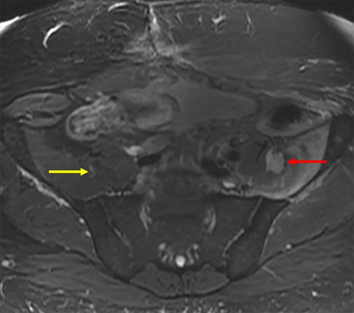
Axial T2-weighted MR image in a 46-year-old male patient with a post–microwave ablation femoral nerve injury. The femoral nerve on the treated side could not be separated from the tumor (red arrow). The location of the femoral nerve on the contralateral side (yellow arrow) is noted for comparison.

Axial T2-weighted MR image in a 46-year-old male patient with a post–microwave ablation femoral nerve injury. The femoral nerve on the treated side could not be separated from the tumor (red arrow). The location of the femoral nerve on the contralateral side (yellow arrow) is noted for comparison.