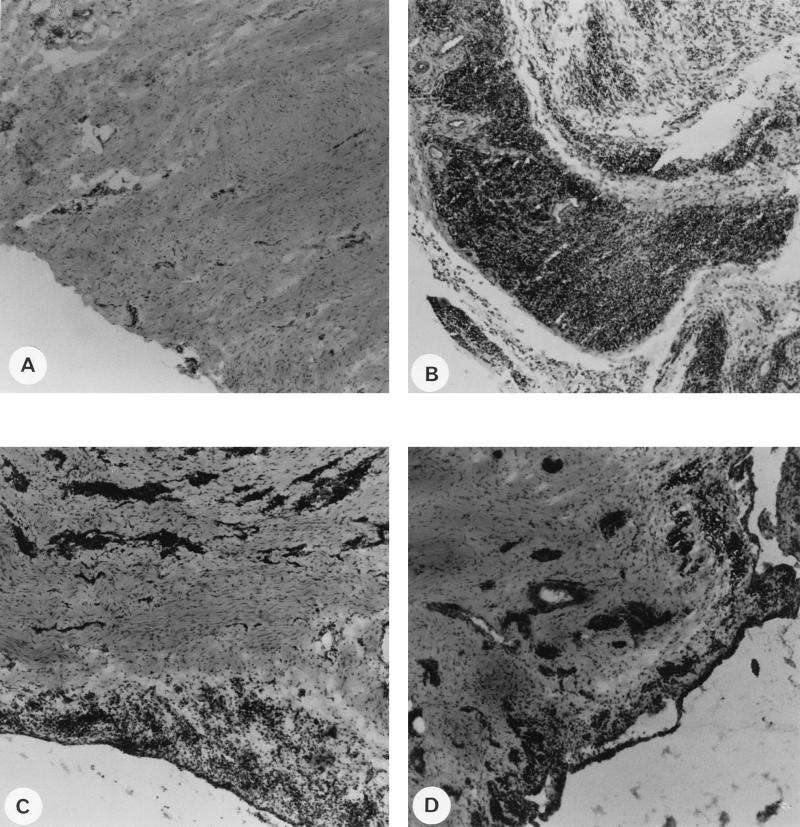
FIG. 4.
Histopathology of synovial membranes from a naive goat (A), from positive control goat 9317, which was injected with CAEV wt (B), from mock-challenged goat 9319, which was inoculated with CAEV tat− (C), and from wt-challenged goat 9327, which was inoculated with CAEV tat− (D). Synovial membrane samples were taken at necropsy, and frozen sections were stained with hematoxylin and eosin. Magnification, ×10.

