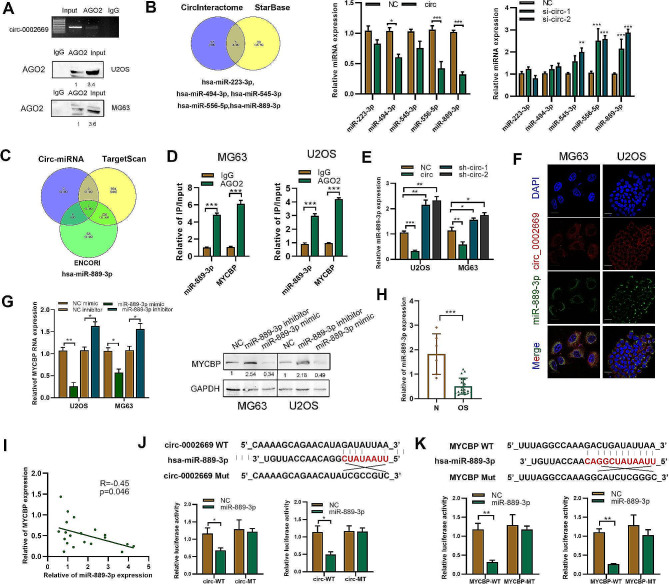
Fig. 4.
Circ_0002669 functions as a sponge for miR-889-3p. B RIP and RT-PCR was performed using U2OS cells to determine the enrichment of circ_0002669. RNA pull-down was performed, followed by western blotting to determine AGO2 expression. B The potential miRNAs binding to circ_0002669, as predicted by StarBase and CircInteractome. Expression of the miRNAs was determined in circ_0002669-overexpressing or -knockdown OS cells. C Identification of miR-889-3p by using TargetScan and ENCORI to screen for upstream miRNAs of MYCBP. D RIP was performed to detect the binding between miR-889-3p or MYCBP mRNA and AGO2 protein. E MiR-889-3p expression was determined using qRT-PCR in circ_0002669-overexpressing or -knockdown OS cells. F FISH was performed to determine the intracellular location of circ_0002669 (red) and miR-889-3p (green) (scale bar, 20 μm). G Expression of MYCBP was determined in miR-889-3p mimic- or inhibitor-transfected OS cells, as determined by qRT-PCR and western blotting. H Relative expression of miR-889-3p in OS tissues (n = 12) and non-tumor tissues (n = 5) was determined by qRT-PCR. I Correlation between miR-889-3p and MYCBP expression in OS tissues. J Schematic illustration of circ_002669-wildtype (WT) and circ_002669-Mut (MT) luciferase reporter vectors. Relative luciferase activities were determined in the indicated transfected OS cells. K Schematic illustration of MYCBP-WT and MYCBP-MT luciferase reporter vectors. The relative luciferase activities were quantitated in the indicated transfected OS cells. Data shown are from three independent experiments, *p < 0.05, **p < 0.01, ***p < 0.001

