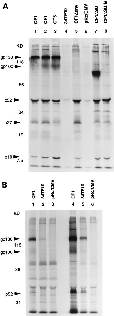
FIG. 2.
Expression and processing of FIV proteins in transfected human and feline cells, assessed by radioimmunoprecipitation with FIV (Petaluma strain)-infected domestic-cat plasma. 293 cells (panel A, all 8 lanes), HeLa (panel B, lanes 1 to 3), and CrFK cells (panel B, lanes 4 to 6) were transfected with 5 μg of the indicated plasmids by calcium phosphate precipitation in 25-cm2 flasks. At 27 h (293 cells) or 44 h (HeLa and CrFK cells) after transfection, cells were radiolabeled and immunoprecipitated (see Materials and Methods). The HeLa cell lysate used for lane 1 in panel B was derived from approximately 15 to 25% of the amount of cells in the other lanes because of the loss of cells to extensive syncytial lysis. The data from one of two radioimmunoprecipitations performed are shown; each yielded the same results. Shown on the left are the positions of molecular mass markers (in kilodaltons).

