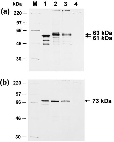
FIG. 3.
Identification of the UL0 and UL[−1] proteins. Radiolabeled proteins were incubated with monospecific anti-UL0 (a) and anti-UL[−1] sera (b) and immunoprecipitated. The in vitro translation products of pRC-UL0 and pRC-UL[−1] (lanes 1) were compared to proteins from primary chicken kidney cells transfected with the respective plasmid (lanes 2). In addition, lysates of chicken kidney cells were analyzed 24 h after infection with ILTV at an MOI of 5 (lanes 3). Results from precipitation of noninfected cell lysates are shown in lanes 4. In the depicted fluorograms of discontinuous SDS–10% polyacrylamide gels, the molecular masses of marker proteins (M) are indicated on the left, and locations of UL0 (61 and 63 kDa) and UL[−1] (73 kDa) gene products are marked by arrows.

