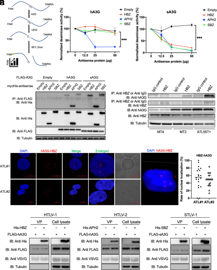
Fig. 2.
Antisense proteins interact with A3G. (A) Schematic representation of the A3G-mediated deaminase activity assay (Left) and quantifications of A3G deaminase activity in the presence of increasing concentrations of HBZ, APH-2, and SBZ (normalized mean ± SD of triplicate experiments; one-way ANOVA with Tukey correction; ***P < 0.001) (Right). (B) Coimmunoprecipitation experiment showing the interaction between human or sA3G and the antisense proteins of HTLV-1, HTLV-2, and STLV-1 in transfected HEK293T cells. IP, immunoprecipitation; IB, immunoblot. (C) Coimmunoprecipitation experiment showing the interaction of endogenous hA3G with endogenous HBZ in HTLV-1-infected T cell lines (MT-2 and MT-4), and an ATL cell line (ATL-55T+). IP, immunoprecipitation; IB, immunoblot. (D) Complex of hA3G with HBZ in primary ATL cells (n = 2; ATL#1 and ATL#2) and 3-dimensional image of a fresh ATL cell (ATL#2) shown by confocal z-stacking, detected by the Duolink Proximity Ligation Assay (Left) and rate of nuclear localization of HBZ-hA3G complex (Right). (E) Immunoblot showing the incorporation of both A3G and antisense proteins into HTLV-1, HTLV-2, and STLV-1 VP. All experiments were performed at least twice.

