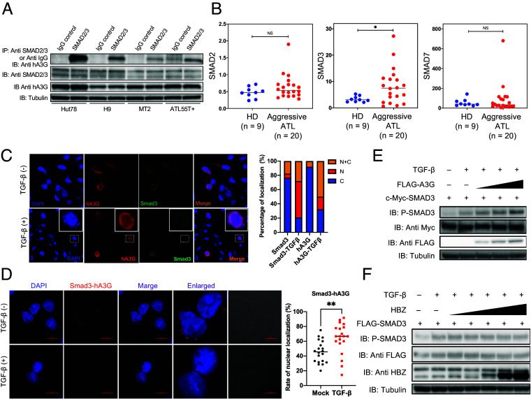
Fig. 4.
Interaction between A3G and Smad proteins. (A) Coimmunoprecipitation experiment showing the interaction of endogenous hA3G with endogenous Smad2/3 in HTLV-1-negative T cell lines (Hut78, H9), an HTLV-1-infected T cell line (MT-2), and an ATL cell line (ATL55T+). IP, immunoprecipitation; IB, immunoblot. (B) Expression of Smad2, Smad3, and Smad7 in healthy donors (n = 9) and patients with aggressive ATL (n = 20) by RT-qPCR (triplicate experiments; one-way ANOVA with Tukey correction; *P < 0.05). (C) Immunofluorescence microscopy images showing the hA3G protein and Smad3 protein in the presence or absence of TGF-β (Left) and percentage of localization (Right) in transfected HeLa cells. N: nucleus, C: cytoplasm. (D) Complex of hA3G with Smad3 in primary ATL cells with or without TGF-β treatment, detected by the Duolink PLA (Left) and the rate of nuclear localization (Right; two-tailed unpaired Student’s t test; **P < 0.01). (E) Immunoblot showing that hA3G expression induces the phosphorylation of Smad3 in a dose-dependent manner in transfected HepG2 cells. (F) Immunoblotting reveals no dose-dependent phosphorylation of Smad3 induced by HBZ expression in transfected HepG2 cells. Experiments were performed at least twice (A and C–F).

