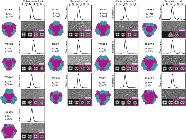
Fig. 2.
Structural characterization of 13 co-expressed ProteinMPNN-designed tetrahedral nanoparticles. Left: Computational design model and the PDB entry from which each of the two trimeric components (A, purple; B, blue) were derived. Top: Co-expressed nanoparticles were analyzed by SEC using a Superdex 200 10/300 column to determine their size and purity. Thirteen out of the 76 designed nanoparticles eluted at ~13 mL, consistent with an expected molecular weight (MW) of ~1 MDa. The absorbance was measured at 230 nm and normalized. Middle: Negatively stained electron micrographs of co-expressed tetrahedral nanoparticles. (Scale bar: 50 nm.) Bottom Left: Two representative 2D projections calculated from the design model. Bottom Right (boxed in purple): corresponding experimentally determined 2D class averages. In all cases, the 2D class averages closely resemble the 2D projections. In the case of T33-11.1, only a single preferred particle orientation was observed experimentally.

