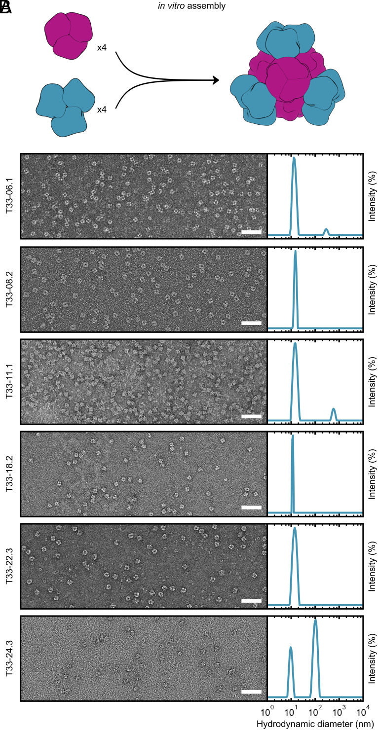
Fig. 4.
In vitro assembly of ProteinMPNN-designed tetrahedral nanoparticles. (A) In vitro assembly of equimolar components A and B at 25 µM in TBS buffer. (B) Left: Negatively stained electron micrographs for 6 ProteinMPNN-designed in vitro assembled nanoparticles. (Scale bar: 50 nm.) Right: DLS of in vitro assembled tetrahedral nanoparticles. A scattering peak centered around the expected diameter (~15 nm) was present for all assembled nanoparticles. For T33-06.1, T33-11.1, and T33-24.3, a second aggregate peak of diameter >100 nm was observed. However, in all instances these peaks represented <1% of the total mass.

