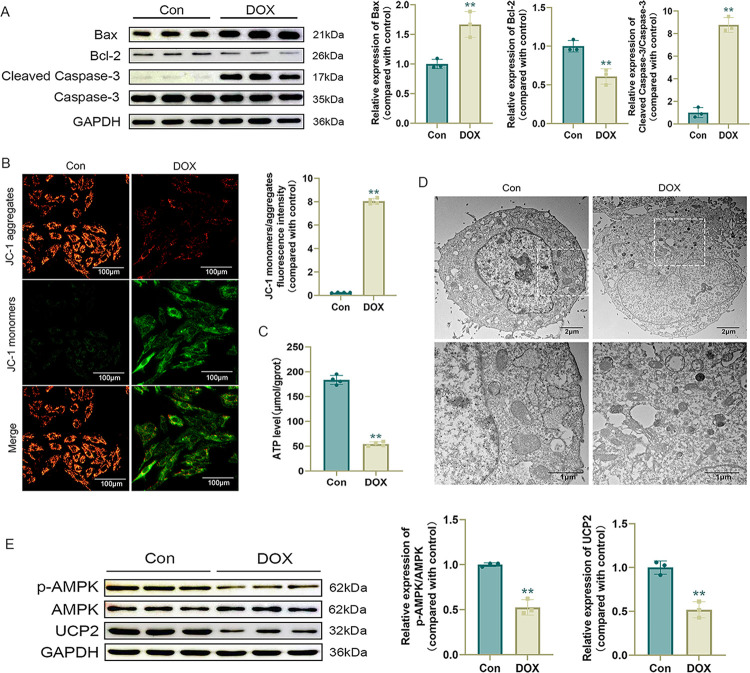
Fig 2. DOX induces mitochondrial damage in H9c2 cells.
(A) Western blot detection of apoptosis-related protein levels and statistical results (n = 3); (B) Representative JC-1 images and quantification of fluorescence intensity for JC-1 monomers/aggregates (n = 4); (C) ATP level (n = 4); (D) Representative images of mitochondria in H9c2 cells observed by transmission electron microscopy; (E) Western blot detection of p-AMPK, AMPK, and UCP2 levels and statistical results (n = 3). Values are presented as the mean ± SD. *p<0.05 vs. Con group, **p<0.01 vs. Con group.

