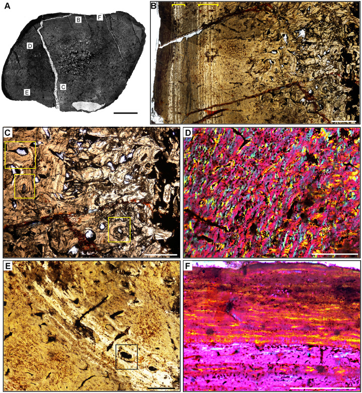
Fig 8. Femur histology of Exaeretodon argentinus PVSJ 38–2002.
(A) General view of femoral histology in plane polarized light. Letters indicate positions of higher magnification photomicrographs B–F. Anterior is toward the top. Scale bar = 5 mm. (B) PPL image of a thickened, compacted cortex (left) that surrounds a small medullary space filled with bony trabeculae that have undergone multiple generations of endosteal remodeling (right). Several cycles of annuli and LAG are visible in the mid- and outer cortex (yellow brackets). The periosteal surface of the element is at the left. Scale bar = 5 mm. (C) PPL image highlighting secondary remodeling. Remodeling is indicated by both erosion rooms and secondary osteons (yellow rectangles), and is confined to perimedullar regions of the deep cortex. The single generation of secondary remodeling leaves persistent patches of primary bone tissue throughout the cross section. Scale bar = 250 microns. (D) XPL with lambda compensator image illustrates the highly vascularized fibrolamellar bone tissue characteristic of most cortical appositional growth in Exaeretodon. Bright pink areas of isotropic bone mineral organization highlight the woven bone component of the fibrolamellar complex. Turquoise and yellow areas reveal the more highly organized, anisotropic lamellar bone mineral organization within primary osteons of the fibrolamellar complex. In this view, primary vasculature is mostly longitudinal, with occasional circular and rare radial anastomoses. Scale bar = 500 microns. (E) PPL image of a broad zone of mid-cortical annuli and LAG. Rare secondary osteons occur between each annulus (black rectangle). Following deposition of these annuli (toward the bottom left corner of this image), appositional growth resumes, but with reduced primary vascularity dominated by mature longitudinal primary osteons. Scale bar = 500 microns. (F) XPL with lambda compensator image of periosteal surface. Here in the outermost cortex, bone tissue is nearly avascular and exhibits a significant increase in bone mineral organization to a lamellar bone matrix. The periosteal surface exhibits multiple stacked Lines of Arrested Growth (LAG) highlighted by yellow in this view. These LAG form the External Fundamental System (EFS) that signals the end of major appositional growth and the attainment of skeletal maturity. Scale bar = 500 microns.

