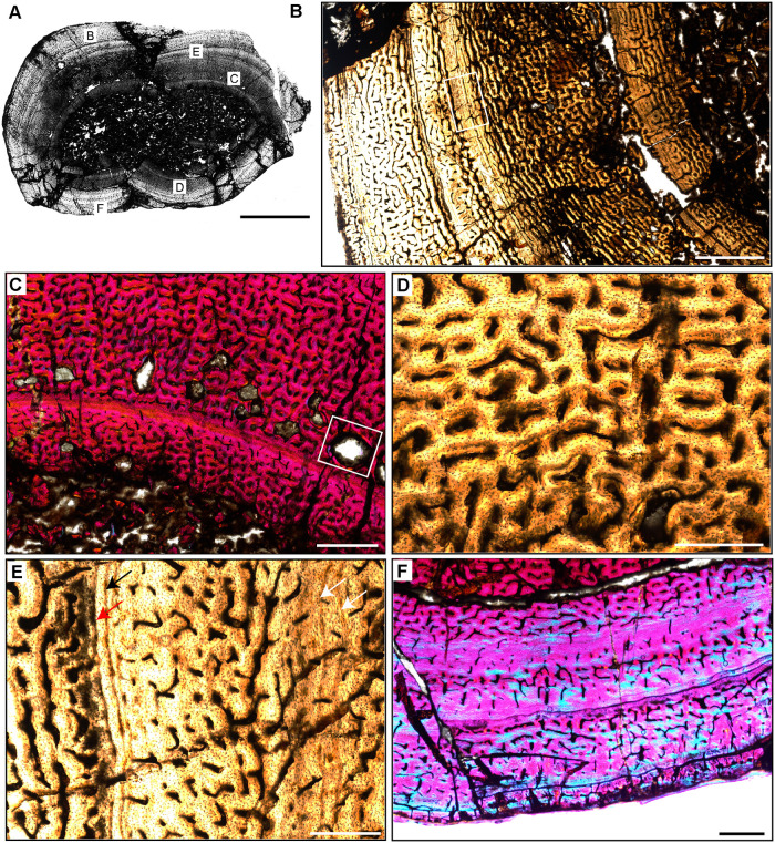
Fig 9. Femoral Histology of Hyperodapedon sanjuanensis PVSJ 574.
(A) General view of femoral histology in plane polarized light (PPL). Letters indicate positions of higher magnification photomicrographs B–F. Anterior is toward the top. Scale bar = 10 mm. (B) PPL image of a thick highly vascularized cortex (left) that surrounds a medullary space that appears to be filled with broken bony trabeculae (right). Primary vascularity is high throughout most of the cross-section, though in the area indicated by the white rectangle a circumferential shift to more organized bone mineral signals a temporary reduction in bone apposition. Subsequently, additional cycles of annuli and LAG are visible nearer the periosteal surface (to the left). Scale bar = 1 mm. (C) XPL with lambda compensator image highlighting the onset of secondary remodeling in the deeper regions of the cortex. Remodeling is indicated by the presence of erosion rooms, a few of which exhibit centripetal deposition of lamellar bone indicating incipient formation of secondary osteons (white rectangle). Signatures of remodeling are mostly confined to the mid-cortex, except for one large erosion room in the outer cortex (seen in A). Scale bar = 1 mm. (D) PPL image highlighting the highly vascularized fibrolamellar bone tissue characteristic of the majority of cortical appositional growth in Hyperodapedon. In this region, primary osteons interweave in a reticular pattern. Scale bar = 500 microns. (E) PPL image of a typical mid-cortical growth cycle in Hyperodapedon. In this image, the deeper cortex is toward the right; the more superficial cortex is toward the left. At the left, faint annuli occur in the context of relatively lower vascularity and osteocyte lacunae that are arranged in parallel layers within a small region of PFB (white arrows). In the middle of this view, vascularity is high, reticular primary osteons are dominate, and occur within a woven bone context to form a typical fibrolamellar complex. Toward the left and more superficially, a reduction in relative bone appositional rate is recorded by a reduction in primary vascularity and a corresponding shift to more organized PFB, followed by deposition of an annulus (black arrow) and a LAG (red arrow). Following deposition of this LAG, the resumption of elevated primary bone depositional rate is heralded by the highly vascularized reticular FLB on the far left. Scale bar = 500 microns. (F) XPL with lambda compensator image from periosteal surface. Here in the outermost cortex, primary vascularity is reduced compared to the deeper and mid-cortex. A transition to more abundant PFB and/or LFB is recorded by the turquoise zones in this image, which indicate the isotropic nature of bone mineral in the outer cortex. The periosteal surface lacks evidence of an EFS. Scale bar = 1 mm.

