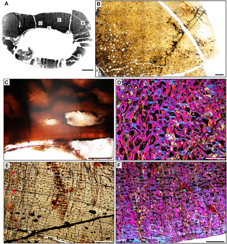
Fig 13. Femoral Histology of Saurosuchus galilei PVSJ 047 (Figs 5C, 5D and 12).
(A) General view of femoral histology in PPL. Letters indicate positions of higher magnification photomicrographs B–F. Anterior is toward the top. Scale bar = 10 mm. (B) PPL image of the highly vascularized remodeled cortex and open medullary cavity in Saurosuchus. The medullary space is at the bottom left; the periosteal surface is at the upper right. The crack visible mid-frame follows a LAG. Even at this low magnification intensive remodeling that extends well into the mid-cortex is visible. Scale bar = 1 mm. (C) PPL view of the perimedullar deep cortex. An IFS overprints two erosion rooms and indicates medullary drift and remodeling in Saurosuchus. (D) XPL with lambda compensator image of the deep cortex highlighting the intensity of secondary remodeling. A single generation of secondary remodeling overprints some areas of primary bone in the deep cortex. Scale bar = 1mm. (E) PPL image of the Saurosuchus mid-cortex. Primary bone tissue is fibrolamellar with a dense laminar vascular network with occasional radial anastomoses. At least six mid-cortical LAG punctuate growth in this specimen; two are indicated here by red arrows. A third deeper LAG traceable in other regions of the cross section is aligned with the crack at the bottom of the frame. Note the erosion rooms and occasional secondary osteons extending into the mid-cortex. Scale bar = 1 mm. (F) XPL with lambda compensator image, outer cortex and periosteal surface. Laminar primary osteons in an FLB context grade into a more organized bone tissue dominated by PFB and patches of LFB with fewer smaller longitudinal primary osteons as we approach the periosteal margin of the bone (bottom). Note that even here in the external cortex a few sparsely distributed secondary osteons exist (white rectangles) and indicate the extent of sub-periosteal cortical bone remodeling. Scale bar = 1 mm.

