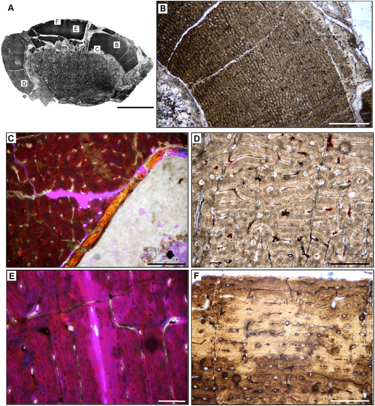
Fig 14. Femoral Histology of Trialestes romeri PVSJ 368.
(A) General view of femoral histology in PPL. Letters indicate positions of higher magnification photomicrographs B–F. Anterior is toward the top. Scale bar = 1 cm. (B) PPL image of cortical bone tissue. Medullary cavity toward bottom left, periosteal surface at top right. Densely interweaving vascular networks characterize Trialestes femoral histology. Scale bar = 300 microns. (C) XPL with lambda compensator image of the deepest cortex. Band of yellow-orange highlights the IFS, a signature of medullary drift and deep cortical remodeling, which cross-cuts densely vascularized primary fibrolamellar bone tissue. Note the absence of other forms of remodeling, including erosion rooms and/or secondary osteons. Scale bar = 300 microns. (D) PPL image of mid-cortex records densely vascularized laminar primary fibrolamellar bone in which circular and longitudinal primary osteons interweave. Scale bar = 300 microns. (E) XPL with lambda compensator image of mid cortex documenting a patch of the superficial primary cortex recording a transition to LFB and/or PFB with a significant, but temporary reduction in vasculature. At least one area of high birefringence resembles a LAG, but it cannot be traced circumferentially around the cross-section, and includes small longitudinal simple vascular canals within. The thickness of adjacent laminae indicate that this birefringence may simply be a circular primary osteon interweaving with a few longitudinal primary osteons within a typical laminar vascular network. Scale bar = 100 microns. (F) PPL image of the external cortex of Trialestes. Deposition of laminar FLB persists, though longitudinal primary osteons become more common than their anastomosing circular primary osteons. At the periosteal surface (top) a deceleration of bone apposition is recorded by a shift toward more organized osteocyte lacunae in parallel rows within an PFB/LFB context, and vasculature becomes dominated by unidirectional longitudinal primary osteons. The EFS is absent, indicating ongoing, but slower growth at this later phase of Trialestes ontogeny. Scale bar = 300 microns.

