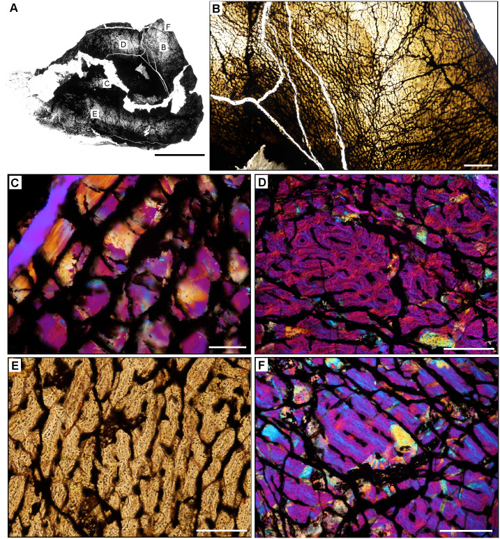
Fig 15. Femoral histology of Sanjuansaurus gordoilloi PVSJ 605.
(A) General view of femoral histology in PPL. Letters indicate positions of higher magnification photomicrographs B–F. Anterior is toward the top. Scale bar = 10 mm. (B) PPL image highlighting brecciated and diagenetically altered preservation of Sanjuansaurus. In spite of relatively poor preservation, primary histological features can still be observed. Sanjuansaurus exhibits an open medullary cavity and a densely vascularized primary bone cortex. Scale bar = 1 mm. (C) XPL image with lambda compensator highlights thin avascular endosteal lamellae surrounding the open medullary cavity (top left) and forming an IFS. The deep cortex lacks other evidence of secondary remodeling. Scale bar = 100 microns. (D) XPL with lambda compensator image of mid-cortex documents fibrolamellar bone tissue that is highly vascularized by reticular primary osteons. Scale bar = 300 microns. (E) PPL image of more superficial cortex indicates the consistency of laminar and/reticular primary osteons in a fibrolamellar context that dominate the appositional growth pattern of Sanjuansaurus. The cortex is completely devoid of secondary osteons, erosion rooms, annuli, and LAG. Scale bar = 300 microns. (F) XPL with lambda compensator image of the external cortex. Periosteal surface is toward top right corner of image. Blue areas highlight more organization of the bone matrix, with osteocyte lacunae organized into parallel lines. In the outer regions of the cortex the woven bone scaffold transitions to a parallel-fibered organization. Primary osteons (here highlighted in pink and orange) are more unidirectional, and usually longitudinal. This femur lacks an EFS. Scale bar = 300 microns.

