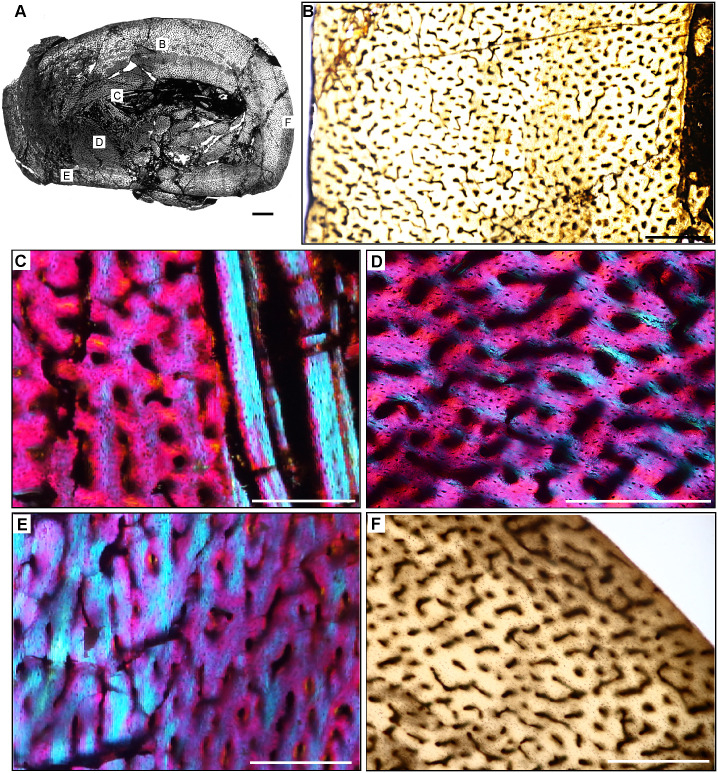
Fig 17. Femoral Histology of Eodromaeus murphi PVSJ 561.
(A) General view of femoral histology in PPL. Letters indicate positions of higher magnification photomicrographs B–F. Anterior is toward the top. Scale bar = 1 mm. (B) PPL image spanning the medullary cavity (right) to the periosteal surface (left). A densely vascularized fibrolamellar cortex devoid of secondary osteons and/or erosion rooms surrounds an open medullary cavity. Scale bar = 500 microns. (C) XPL with lambda compensator image details lamellar fibered layers of endosteally derived bone that form an IFS lining the medullary cavity (turquoise at right). Primary tissue in the deep cortex overprinted by the IFS is reticular fibrolamellar bone, seen here in pink with orange and turquoise illustrating lamellar bone organization surrounding primary vascular canals. Scale bar = 250 microns. (D) XPL with lambda compensator image drawn from mid-cortex illustrates osteonal organization of lamellar fibered tissue around primary vascular canals within a woven bone scaffold. Note the reticular organization of primary vascular canals and the rounded and numerous osteocyte lacunae. Scale bar = 250 microns. (E) XPL with lambda compensator image near the periosteal surface highlights primary bone organizational change in later ontogeny. The middle cortex (toward the right) records a traditional fibrolamellar complex with reticular primary osteons indicative of relatively elevated growth rates. Closer to the periosteal surface (toward the left) the scaffold of apposition transitions to parallel-fibered bone mineral, indicated by the turquoise color in this micrograph. Scale bar = 250 microns. (F) PPL image of the external cortex highlighting an area of ongoing but slowed growth at the periosteal surface. Primary reticular fibrolamellar bone persists more deeply, but at the periosteal border osteocyte lacunae become more organized in parallel layers reflecting the parallel organization of bone mineral. These changes are accompanied by a shift toward more sparse longitudinal vascular canals. Together, these changes indicate a reduction in appositional growth but not a cessation. An EFS is absent in Eodromaeus. Scale bar = 200 microns.

