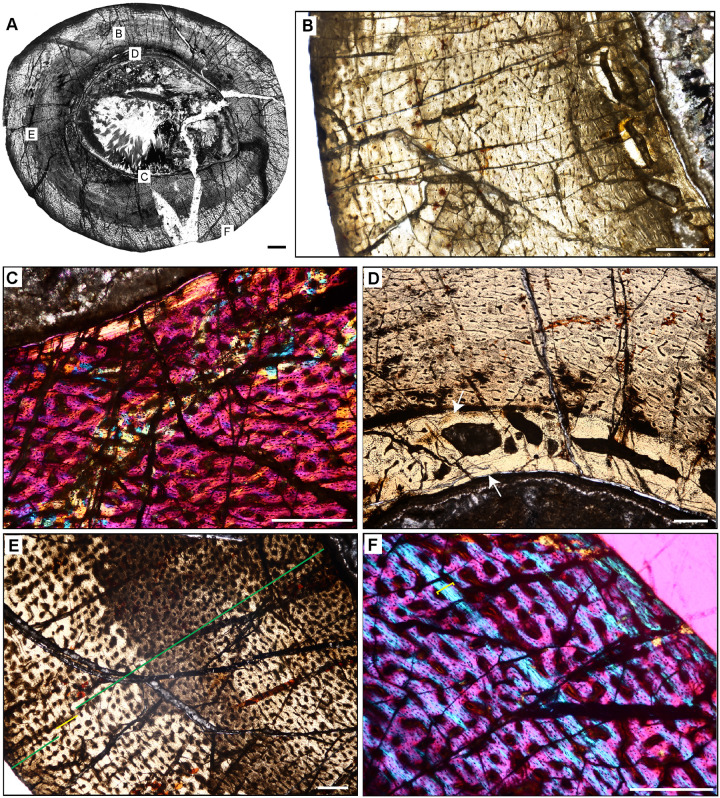
Fig 18. Femoral histology of Eoraptor lunensis PVSJ 559.
(A) General view of femoral histology in PPL. Letters indicate positions of higher magnification photomicrographs B–F. Anterior is toward the top. Scale bar = 1 mm. (B) PPL image spanning the medullary cavity (right) to the periosteal surface (left). Unusually large erosional spaces are visible in the deep cortex. The cortex preserves well-vascularized fibrolamellar bone. LAG are absent. Scale bar = 500 microns. (C) XPL with lambda compensator image of perimedullar cortex. Orange color in the top left highlights the well-developed IFS lining the medullar cavity. The deep cortex exhibits fibrolamellar bone dominated by abundant longitudinal primary osteons. Scale bar = 300 microns. (D) PPL image of perimedullar erosion bays that are situated between two layers of endosteally-derived lamellae (white arrows) that have eroded primary bone tissue and reflect medullary drift. Between these thin lamellar layers large erosion rooms have eroded away compacted Haversian osteonal bone. Scale bar = 300 microns. (E) PPL image of the cortex Eoraptor documenting three observable growth intervals, in the context of continuous deposition of well-vascularized fibrolamellar bone. The deepest cortex (green line, upper right) exhibits abundant circular and longitudinal primary osteons that sometimes interweave in a laminar pattern. This segment encompasses the majority of appositional growth in Eoraptor. Following this burst of growth, a period of deceleration is indicated by a shift toward less primary vasculature and parallel-fibered bone tissue (yellow line). A return to faster growth later in ontogeny is indicated by more highly vascularized fibrolamellar bone (green line, lower left). In some regions of the last growth interval at the periosteal surface circular, longitudinal, and radial primary osteons interweave in a sub-plexiform pattern. Scale bar = 300 microns. (F) XPL with lambda compensator image of another area of the outer cortex in Eoraptor. This image highlights the same pattern observed in (E, yellow line) zooming in on the region of growth deceleration. Parallel-fibered bone tissue surrounds mostly longitudinal primary osteons (lower left). The bright turquoise line (yellow bracket) indicates a transition to avascular lamellar fibered bone tissue signaling a temporary but significant decrease in bone apposition. This annulus is not traceable circumferentially. More periosteally, primary osteons exist in a woven-fibered scaffold to form a traditional, highly vascularized fibrolamellar complex during the final recorded phase of growth for Eoraptor. The lack of an EFS indicates that this individual was still actively growing at the time of death. Scale bar = 300 microns.

