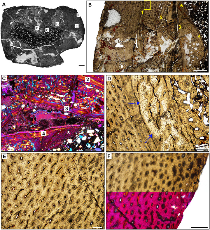
Fig 19. Femoral histology of Chromogisaurus novasi PVSJ 845.
(A) General view of femoral histology in PPL. Letters indicate positions of higher magnification photomicrographs B–F. Anterior is toward the top. Scale bar = 1 mm. (B) PPL image spanning the medullary cavity (right) to the periosteal surface (left). Four cycles of bone remodeling each include centrifugal erosion, followed by centripetal deposition of endosteally-derived LFB, and continued erosion of this LFB through the formation of secondary osteons/dense Haversian bone tissue. The endosteal LFB layers for each of these cycles is indicated by the numbers, 1–4, with 1 representing the initial erosional cycle, and 4 representing the cycle that was most close to the time of death/most recent. The same numbers and the star also apply to cycles labeled in C. The yellow rectangle indicates the approximate position of D. Note the unusually large erosional spaces are visible deep to cycle 3, and in the patch of primary woven-fibered bone tissue lining the perimedullar space, indicated by the yellow star. Superficial to the most external layer of endosteal lamellae the cortex preserves well-vascularized fibrolamellar bone. LAG are present but only in association with deep cortical remodeling. Scale bar = 500 microns. (C) XPL with lambda compensator image of perimedullar cortex. Numbers indicate the intervals of deep cortical centripetal deposition of lamellar bone tissue following an episodic cycle of medullary expansion. Between these intervals, these secondary bone tissue deposits are also eroded, and are replaced by secondary osteons that form dense Haversian bone. This signature is especially clear between Cycles 2 and 3 in this image. Cycle 4 includes both endosteally-derived LFB, secondary osteonal bone, and longitudinally vascularized woven fibered bone that lines the medullary cavity (indicated by the white star). Scale bar = 300 microns. (D) PPL image of perimedullar secondary osteons and erosion rooms are situated between two layers of endosteally-derived lamellae (cycles 1 and 2 in image B). Blue arrows indicate the superficial extent of centrifugal erosion and reversal in cycle 1. Secondary osteons gradually replace earlier deposits of endosteally-derived LFB. More deeply, between cycles 1 and 2, secondary osteons have obliterated primary bone tissue. Scale bar = 500 microns. (E) PPL image of the cortex Chromogisaurus documenting the general nature of the middle cortex. Superficial to deep cortical signatures of remodeling, primary fibrolamellar bone tissue vascularized by abundant longitudinal vascular canals persists to the external cortex. In some regions of the mid-cortex circular and longitudinal primary osteons anastomose in a sub-laminar to laminar vascular arrangement. Scale bar = 250 microns. (F) PPL and XPL with lambda compensator composite image of another area of the outer cortex. Deposition of fibrolamellar bone tissue vascularized by longitudinal primary osteons persists in preserved regions of the external cortex. The lack of an EFS or a significant change in bone depositional pattern indicates ongoing appositional growth in Chromogisaurus at the time of death. Scale bar = 500 microns.

