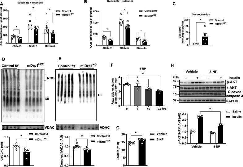
Fig. 4. Drp1 deletion reduces mitochondrial Complex II assembly and activity in muscle from male mice.
(A) Oxygen consumption rate (OCR) of mitochondria isolated from gastrocnemius muscles of NC-fed control f/f and mDrp1HET mice, and (B) mitochondria from control f/f and miDrp1KO mice treated with the succinate and rotenone (n = 5 per genotype). (C) Succinate levels in skeletal muscle of NC-fed control f/f and mDrp1HET mice (n = 5 per genotype, 5 months of age). (D) Representative Blue Native polyacrylamide gel electrophoresis (BN-PAGE) gels showing complex II assembly (blot for SDHA) of the gastrocnemius muscles of control f/f and mDrp1HET mice, and (E) muscle from control f/f and miDrp1KO mice (n = 5 per genotype). (F) Fatty acid oxidation of C2C12 myotubes with 3-NP administration (n = 3 biological replicates). (G) Lactate levels in the culture medium of differentiated control (Scr) and Drp1KD myotubes treated with 3-NP (100 μM, 24 hours). (H) Immunoblot of p-Akt serine-473, total Akt, and cleaved caspase 3 in C2C12 myotubes incubated with vehicle or 3-NP (complex II inhibitor, 100 μM, 24 hours) with and without insulin (10 nM, 15 min) (n = 3 biological replicates). All values are presented as means ± SEM; *P < 0.05 determined by unpaired Student’s t test, two-tailed.

