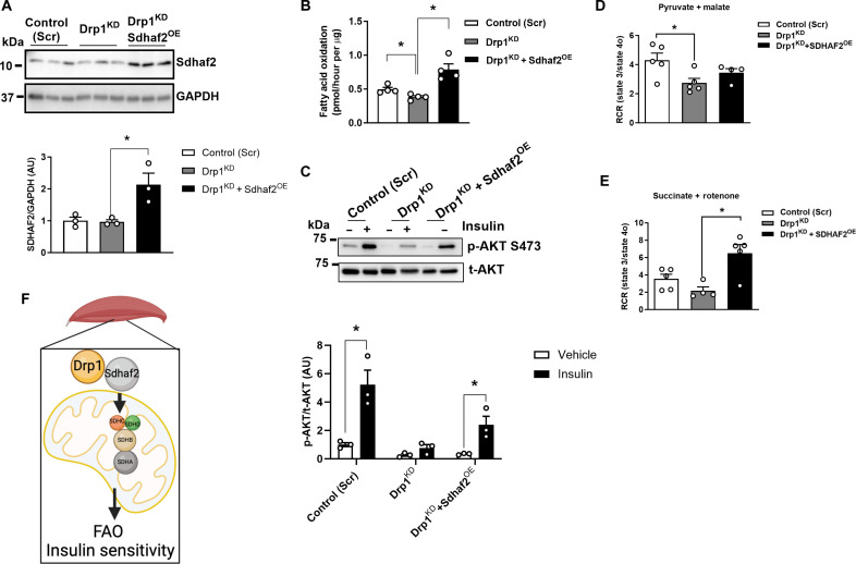
Fig. 7. Sdhaf2 overexpression restores fatty acid oxidation and insulin action in DrpKD myocytes.
(A) Immunoblot of Sdhaf2 in control (Scr), Drp1KD, and Drp1KD and Sdhaf2OE myocytes. Densitometric quantification of Sdhaf2 protein normalized to GAPDH (n = 3 biological replications). (B) Fatty acid oxidation analysis of control (Scr), Drp1KD, and Drp1KD and Sdhaf2OE myocytes (n = 4 biological replicates). (C) Immunoblot of phospho-Akt serine-473 and total-Akt in control (Scr), Drp1KD, and Drp1KD and Sdhaf2OE myotubes with and without insulin (n = 3 biological replications). Respiratory control ratio (RCR) of control (Scr) versus Drp1KD versus Drp1KD and Sdhaf2OE myocytes with (D) pyruvate and malate or (E) succinate and rotenone (n = 4 to 5 per genotype). (F) Schematic overview of Drp1-Sdhaf2 interaction promoting mitochondrial translocation to enhance fatty acid oxidation (FAO) and insulin sensitivity in skeletal muscle. All values are presented as means ± SEM; *P < 0.05 determined by unpaired Student’s t test, two-tailed.

