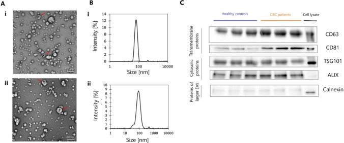
Fig. 1.
Characterization of isolated sEVs. A Representative transmission electron microscopy images of serum-derived sEVs isolated using size exclusion chromatography (scale bar, 200 nm) from CRC patient (i) and healthy control samples (ii). B Representative graphs by DLS analysis indicating concentration and size distribution of isolated particles from CRC patient (i) and healthy control samples (ii). C Representative Western blot images showing enrichment of EV markers CD63, CD81, TSG101, and Alix with a low level of endoplasmic reticulum proteins (calnexin) in serum-derived sEVs

