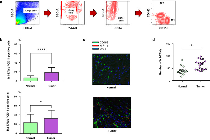
Fig. 1.
The number of M2-TAMs increased in the tumor. a The gating method of flow cytometry for freshly resected surgical specimens to detect M1-TAMs (CD14+CD11c+CD163−) and M2-TAMs (CD14+CD11c−CD163+). b The summary of population of M1- or M2-TAMs in CD14-positive cells in the normal mucosa (normal) and the tumor (tumor) by flow cytometric analysis (M1-TAMs; CD14+CD11c+CD163−, M2-TAMs; CD14+CD11c−CD163+). c Representative images showing the immunofluorescence staining of M2-TAMs in the normal mucosa (normal) and the tumor (tumor) samples. Green staining; CD163, red staining; HIF-1α, blue staining; DAPI. d The number of M2-TAMs in the normal mucosa (normal) and the tumor (tumor). *p < 0.05, ****p < 0.0001

