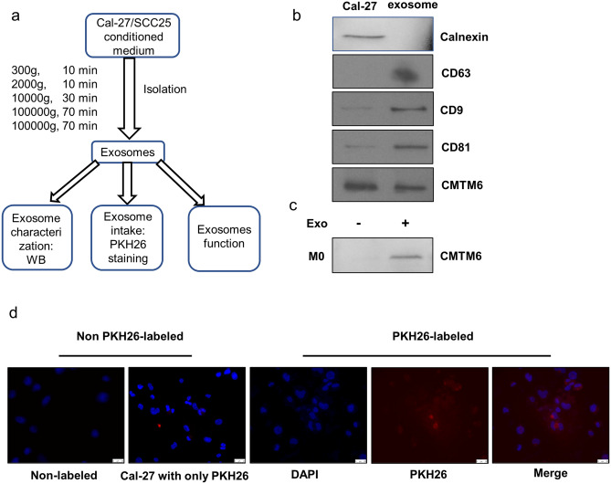
Fig. 4 .
The isolation and identification of Cal-27 exosomes. a A sketch of isolation path and identification of exosomes. b Western blot analyzed of exosomes for CD63, CD9 and CD81 (exosome biomarker), Calnexin (negative marker) and CMTM6. c Western blot analyzed of M0 cells and exosomes incubated M0 cells for CMTM6. d Representative fluorescent images for PKH26-labeled exosomes taken by M0 after 12 h incubation. Exosomes were stained red and M0 nuclei were stained blue by DAPI (scale bar: 25um). Cal-27 cells incubated with non-labeled exosomes (PBS was added to 25 μg exosomes instead of PKH26) and Cal-27 cells with only PKH26 (PKH26 was added to PBS and was re-isolated as stated) were used as controls

